The 2009 Nobel Prize in Chemistry was awarded to Venkatraman Ramakrishnan, Thomas Steitz and Ada Yonath for their work on the structure and function of ribosome, a component of the cell that translates genetic information and synthesizes protein. One of the laureates Prof. Ada Yonath of Israel was recognized for her many years of studies of purification and crystallization of ribosome crucial to the structural analyses. She in the process had also visited KEK for many times to conduct her research at the Photon Factory.
The genetic information of living system is coded in DNA. Living organisms pass on the DNA information from generation to generation to continue their existence. The essential to the biological processes, however, are not the DNAs themselves, but rather proteins. In the work of all organisms, the genetic information is first transcribed to RNA, which is then translated into proteins. These proteins are responsible for various biological processes within the living organisms.
|
 |

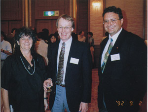
Prof. Ada Yonath (left) at the banquette during BSR92 (the forth International Conference on Biophysics and Synchrotron Radiation) with Prof. Wayne Hendrickson (center) of Colombia University and Dr. Joseph Zaccai (right) of Institut Laue-Langevin.
|
The mechanism of transcription and translation of genetic information describes how the DNA information is converted to proteins that play vital roles in biological system. The process this mechanism describes is crucial and of fundamental nature. Prof. Roger Kornberg was awarded the 2006 Nobel Prize in Chemistry for his work on RNA synthetase that plays a central role in transcribing the genetic information.
For the translation of RNA to proteins, on the other hand, it had been long known that ribosomes were responsible. Ribosomes are very large complexes of multiple types of proteins and RNAs. They translate genetic information of messenger RNA (mRNA) to build long chain of amino acids (protein). The amino acids are delivered by transfer RNA (tRNA) molecules, which read the mRNA instruction and bind in the correct sequence to create proteins. The molecular machinery of the binding and structure of ribosomes, however, had been the long-standing mysteries in protein synthesis.
To reveal the structure of ribosome, several research groups worldwide had launched projects to conduct crystal structure analysis using a method called X-ray crystallography. However, the number of atoms they needed to map for ribosomes is simply too large: a few orders of magnitude greater than other types of protein. The teams were faced with difficulties in both the purification, crystallization and refinement processes.
Prof. Yonath was one who had worked tenaciously on the studies of ribosome crystallization for long years. In her quest, KEK's Photon Factory played a great role.
The field of protein crystal structure analysis now attracts the largest number of users in the Photon Factory at KEK. In 1987, when the first users applications at BL6A, the protein crystal structure analysis station, were accepted, there were only three proposals that were approved. One of them was Prof. Yonath's proposal titled: 'Crystal structure analysis of ribosomal particles from bacterial sources'.
"We were surprised and delighted about the proposal, because we did not expect international applicants from the start," says Professor Emeritus Noriyoshi Sakabe of KEK. Sakabe, the leader of BL6A, designed the large Weissenberg camera that was the core component of the station. He remembers considerable number of packages arriving just before the group led by Prof. Yonath showed up.
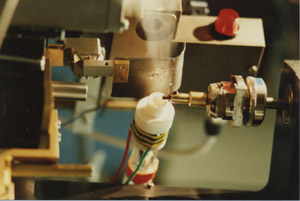
Inside those packages were many pieces of equipment including the glass-made low-temperature nitrogen gas spraying apparatus (see photo). "Back in 1987, Ada
already recognized the key aspect of the X-ray
diffraction experiment," Sakabe says. "Collecting
diffraction data from a large protein molecule like
ribosome had been a very difficult process. What she
did was to establish a method of quickly freeze ribosome
crystal without surrounding water molecules, and to check
the quality of the sample in minutes."
Prof. Yonath diligently immersed a crystal into oil one by one,
froze it with liquid propane, picked it up with
a small size loop made out of human hair, kept its
temperature below -200 deg C with a gas of cooled
nitrogen, exposed it to X-ray and obtained a Polaroid image to survey a good crystal. "She looked tireless when she
repeated the process over and over, until she got
convincing data," Sakabe recalls.

Cryocrystallography system using nitrogen gas below -200 deg C stream developed by Prof. Yonath and her group. (Image: Noriyoshi Sakabe)
|
 |
Her first visit unfortunately did not yield her expected results. Prof. Yonath however did not look at all disappointed. She told Sakabe that she dearly liked the experimental station, and that she would write another proposal very soon because she was expecting good outcome if only she could make the oil and crystal compatible. Then she left joyfully.
For nearly ten years since then, Prof. Yonath and her group came back to
the Photon Factory many times to use Prof. Sakabe's Weissenberg camera
for data collection. After the period, the group continued their work on ribosomes at the
DESY (German Electron Synchrotron) in Hamburg, Germany, to use Doris
beamlines, and the ESRF (European Synchrotron Radiation Facility) in
Grenoble, France, to use their new beamlines including the ones
developed by Prof. Soichi Wakatsuki of KEK (now Director of the Photon
Factory) and others. All the while, the group continued their quest for the structure of ribosomal complexes.
|
In 1992 when Prof. Yonath was still working at the KEK Photon Factory, she gave a special talk on the low-temperature ribosome crystal structure analysis at the forth International Conference on Biophysics and Synchrotron Radiation held in Tsukuba, Japan (see photo).
Our heartfelt congratulations to Prof. Yonath for being awarded the Nobel Prize in Chemistry!
|


