|
|
||||||
| >Top >Feature Story >this page | last update:
10/01/26
|
|
| Watching light-induced molecular dynamics A team at KEK’s Photon Factory Advanced Ring (PF-AR) has been observing the dynamics of various materials excited by light with their new time-resolved X-ray beamline. This growing technique can have great impact on a broad range of fields from environmental science and materials science to biological sciences. |
|
||||||||||||||
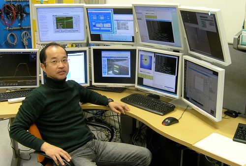
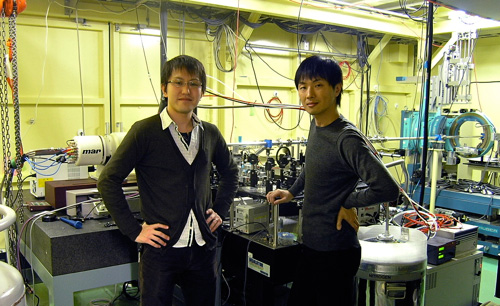
Hop in a submarine, have yourself shrunk to the size of a single cell, and dive into a man's blood stream to save his life. Yes, this is the synopsis from Fantastic Voyage, a1966 film from the U.S. During their rescue mission, the voyagers would witness the beautiful functions of life at work. Prof. Shin-ichi Adachi of KEK admits that just such an adventurous idea might have been the driving force behind his desire to see the dynamics of tiny structures. In reality, life pounds and breathes in rhythms. It is not static like a photo. “But of course, if you are shrunk to nano-scale, you will be smashed before you know it because of the rapid nano-scale motion of molecules, called Brownian motion.” To observe such ultrafast processes in ultrafine structures, the tool Adachi had in mind six years ago when he joined KEK was called time-resolved X-ray structural analysis beamline. X-ray structural analysis is a technique to resolve the structure of materials by using X-ray. X-ray is an electromagnetic wave that has wavelength ranging from 0.01 to 10 nanometers. Since atomic distance in molecule is on the order of 0.1 nanometers, X-ray is perfect for observing such fine structures that optical microscope cannot probe. The mainstream of X-ray structural analysis has been to illuminate samples with a continuous stream of X-ray to get clear pictures of static structures. Adachi’s interest had always been to capture the movements of nano-scale structures. “We function because proteins do work for us, and plants create energy through photosynthesis. We know components in the cell level from static observation, but if we actually observe these processes in motion, we might find something new.”
Adachi had previously been a user of time-resolved X-ray beamline at the European Synchrotron Radiation Facility (ESRF) in France. He had tried to develop a new project for such science when he was at SPring-8, a synchrotron radiation facility in western part of Japan. “The problem was always the short amount of beam time allotted for the time-resolved X-ray experiment, typically two weeks annually,” says Adachi. Thus, he saw an opportunity when he learned that KEK was looking for a useful way of utilizing its Photon Factory Advanced Ring (PF-AR). The Advanced Ring was once a booster for TRISTAN—the precursor of KEK’s main ring accelerator, KEKB. When TRISTAN was shut down for good, AR became the full-time accelerator for materials science. So it is old. Old, but designed to operate in single-bunch mode. This means that only one bunch of electrons goes around AR at a time, producing X-ray pulses only once per 1.3-microsecond cycle. A three-centimeter long electron bunch corresponds to the pulse width of 100 picoseconds (100 x 10-9 seconds). Thus, the AR was a perfect fit for Adachi’s time-resolved experiment. Spectroscopy, diffraction, and scattering Adachi’s plan was to construct a beam line that is dedicated to time-resolved X-ray structural studies. Three years of design and construction and two years of data collection have brought Adachi’s team to their present success. They are now producing impressive results, directly observing a range of phenomena in nano-scale structures. Important current subjects include proteins breathing oxygen in and out and magnetic phase transitions in iron and cobalt compounds.
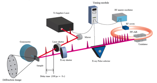
The spectroscopy technique is useful when scientists want to get information about structures in relatively local regions—at the atomic level—of their sample. Diffraction is for when they want to get information about a broader region—often times an entire molecule or complex. The scattering technique has less spatial resolution than the diffraction technique, but is useful in non-periodic structures such as those found in liquids. “We wanted to build a beam line that could provide our users with the ability to use all available techniques,” says Adachi. Their beam line is currently the only beamline in the world that is dedicated full-time to time-resolved X-ray structural studies, and allows users choose from all three techniques. Pump and probe The team is particularly interested in light-induced processes. They first induce some reaction in their samples by exposing the sample to light of a particular color, and then send a 100-picosecond X-ray pulse to observe the process at a single moment in time. By adjusting the timing of laser pulse, they can image the reaction process at different timings. Stringing these images together in sequence, researchers can produce the equivalent of a movie, allowing them to watch the process unfold. This method—excite and observe—is referred to as the pump-probe technique.
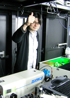
Dr. Tokushi Sato of KEK designed and built the laser system. He set up devices to synchronize the timing of the two experimental processes, to chop off X-ray pulses so as to match the frequency of the pulses to the frequency of laser system, and then to send each laser pulse at well-characterized timing. “It took us three full years to build a successful laser system,” says Sato. He produces laser pulses for the team members and users. The biggest challenge came from the versatile nature of the beamline: the range of laser properties that would be required by users of the system. “Every experiment requires different intensity, wavelength, and pulse width of laser,” explains Sato. “Some experiments need a destructively strong laser, and others need low intensity pulses. The wavelength and pulse width also need to correspond to experiment-specific conditions.” Sato handles three different types of laser systems to produce various types of pulses depending on the subjects. The right place at the right time Creating a new device always requires overcoming challenges. Even after construction is complete, it takes time for researchers to tune and calibrate the system. Adachi says that it took them approximately a year before they were able to collect any meaningful data.
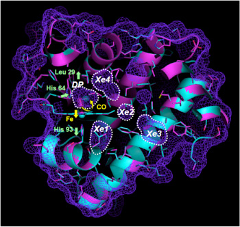
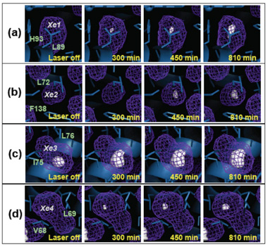
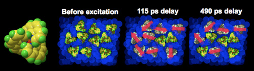
Months later, after trying everything they could think of, the team changed their strategy. The problem was that this first sample was an opaque solid, meaning that only the surface of the sample could be excited by lasers. Because only a small portion of the sample was affected by the light, the team was having a hard time focusing the system on this affected region. So they tried a different type of sample, a material composed of cadmium sulfide whose structure is compressed by light. In this material, the light-induced shock compression penetrates deep inside the material, even to the degree that the sample would be destroyed. “We could immediately see the effect with our setup,” says Adachi. After this, the team rapidly acquired a feel for the nano-scale experiment. Proteins that breathe With the working experimental setup, Dr. Ayana Tomita of the Tokyo Institute of Technology looked at a type of oxygen-carrier protein found in muscle tissue called myoglobin. The myoglobin structure contains many cavities inside. When a carbon monoxide molecule (CO) is introduced to a myoglobin, it binds with iron-based compound called heme inside protein matrix. A pulse of laser light can induce chemical breakdown of the binding, setting the CO molecule free. Those molecules then move around among neighboring cavities. However, actual motion of those molecules through cavities had not previously been observed.
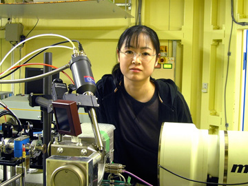
To observe proteins, researchers generally use the diffraction technique because it allows them to gain detailed information about the entire sample. Because Tomita had to take multiple shots per sample to understand how CO molecules moved through the structure, the amount of data quickly grew large. She says that the real challenge was to make the structural analysis of the tens of data sets from each sample. Because the experiment is relatively new, there were no established procedures for analysis. She had to use a different software for each of the dozen steps of her analysis, to ultimately create movies of breathing myoglobin molecules from the electron density distribution data. “Proteins are especially intriguing to me because of their complex structure,” says Tomita. “The time-resolved X-ray structural analysis of proteins is new. I am interested to see how other proteins behave.” Tomita is also interested in rhodopsin, a photosensitive protein found in the retina, and in the proteins at work in photosynthetic processes.
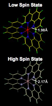
Sub-nanosecond magnets Dr. Shunsuke Nozawa of KEK had been interested in seeing the dynamics of fine structures for long time, and is one of the first members to join Adachi’s group. Nozawa recently succeeded in directly observing the change of magnetism in a molecular complex using the X-ray absorption spectroscopy technique. We know that a solution of iron based molecular complex called iron-phenanthroline undergoes a color change only for a short period of time when the molecular complex is excited by pulsed laser. The color change comes from the change in electronic state of the molecule, which is driven by the change of magnetism. A pulse of laser light changes the spin of the iron atom in this molecular complex by transferring electrons from the iron atom to the ligand atoms that bind to the iron atom. This changes the magnetism in the molecular complex for a very brief 700 picoseconds. Using the absorption technique, Nozawa observed not only the expected change in spin state but also a change in the molecular structure of the complex. The distance between the iron and the nitrogen in the phenanthroline increased by about ten percent. The dynamics are theoretically well explained, but this was the first time it had been directly observed. “What’s interesting about the light-induced ultrafast phenomena is that we can control the function of materials using light,” says Nozawa. “Using pulsed X-ray photons, we can observe ultrafast light-induced changes in the function and structure simultaneously. This is crucial information for molecular design.”
The iron and cobalt based complex undergoes a phase transition when its temperature is increased, and altering the structure of the complex so that it becomes a magnet. This phase transition can also be induced by light. “In the thermal phase transition, the thermal energy built up within the complex induces the change all at once, so it is hard to observe how one-site excitation expands into a macroscopic phase transition,” says Nozawa. “When we induce it using light, we can see how the functional and structural changes propagate through the system via interactions in the crystal.” The light-induced effect is also much quicker than the thermal-induced effect. For this reason, light-induced ultrafast phenomenon makes a good candidate for the future ultrafast devices. Understanding the dynamics of the structure, therefore, is of practical importance. The world’s foremost center for time-resolved X-ray experiments Adachi hopes that, over the next few years, the PF-AR beamline will become the global center for time-resolved X-ray science. The beamline is well on its way to meeting this goal. Already, users from Brazil, Denmark, France, Italy, Korea, UK, US and several national institutes visit Adachi's beam line to conduct their experiments. His team runs the beam line day and night, and supports users 24 hours a day with just handful of members, so as not to waste precious beam time. The team members also have their own plans. Dr. Manabu Hoshino of the Tokyo Institute of Technology joined last April to conduct an experiment that can only be done at the KEK PF-AR beam line. He works on photocatalytic molecules that accelerate photoreactions. Though three-dimensional knowledge of the molecular structure is crucial to understand and control these functions, it is currently unknown. The problem is that the electron excitations due to light last for only short time, on the order of nanoseconds to microseconds. “So far, we have results from simulation and simple spectroscopic analysis,” says Hoshino. “I believe the time-resolved three dimensional analysis will shed new light on our dynamic studies.” He just finished data taking and is now starting the analysis.
In addition to his work on the laser systems, Sato wishes to proceed with his research on energy and environmental technology. In particular, he plans to look into a new type of high-efficiency, inexpensive solar cell called a dye-sensitized solar cell (DSSC). “If we can understand the property of energy conversion, we can apply it to light emission as well,” says Sato. He says he would like to also study a new display technology called organic light emitting diode (OLED). “To understand energy conversion in DSSC and emission in OLED requires good understanding of excited states. For that, we need to study dynamics of the electronic states and structures. The time-resolved X-ray is a powerful tool to investigate the dynamics.” According to Adachi, the beam line’s abilities aren’t that much different from others, but the capacity is much greater, as they have 4,000 hours of beam time per year. “The importance of time-resolved X-ray structural study has gradually increased, with the increasing availability of beam time globally,” says Adachi. “However, to really achieve the science we would like, we need higher quality beam lines in the future.” He adds that this is also a step forward towards the next generation Photon Factory, the energy recovery linac (ERL), planned at KEK. |
| copyright (c) 2010, HIGH ENERGY ACCELERATOR RESEARCH ORGANIZATION, KEK 1-1 Oho, Tsukuba, Ibaraki 305-0801 Japan |
||