While high energy particle accelerators have been the major tool for nuclear and elementary particle researches, electron/positron accelerator rings have taken an other important role to provide a very powerful radiation (called as synchrotron radiation : SR ). KEK Photon Factory (PF) has two electron (positron) accelerator rings dedicated for generating the SR. The first ring (2.5GeV) has been in operation since 1982 and the second ring (6.5 GeV) since 1986. SR is emitted along tangent directions when trajectory of electrons traveling near the speed of light are bent under a magnetic field. It has very unique properties such as (1) high brightness (several orders of magnitude brighter than conventional X-ray sources), (2) a continuous spectrum covering from infrared to X-rays, (3) linearly polarized and (4) pulse width of sub-nanoseconds. Because of these, SR has been used as a tool for observing positions, motions and behaviors of atoms and electrons in various kinds of materials.
Synchrotron Radiation as a microscopic probe
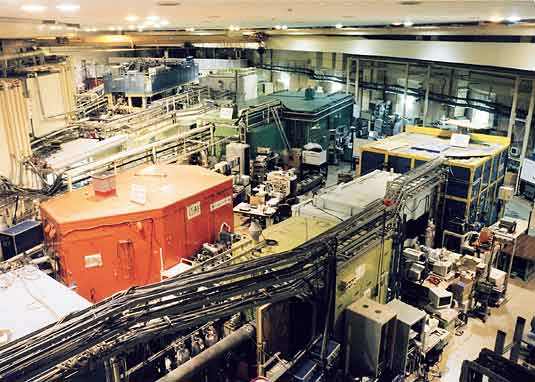
Since SR is a microscopic probe with a resolution to see the size of atoms and molecules, area of its application is very broad such as in physics, chemistry, materials science, biology, medical science, pharmaceutical science/engineering, electronics and so forth. The technique used in those studies are also full of varieties: diffraction, scattering, absorption, fluorescence, secondary emission, radiation effects. To accommodate wide variety of researches, we have about 60 experimental stations. The first picture shows a view of beamlines and experimental stations in 2.5 GeV ring experimental hall.
More than 2,000 researchers visit and stay at the Photon Factory to conduct their experiments to determine precise structure of proteins, viruses, high-temperature superconductors, materials under high pressure and high temperature (to find out what is happening in the inner core of the earth) and to study radiation-induced chemical reactions, atomic level mechanism of catalytic reaction, atomic level analysis of the mechanism of muscle contraction, trace element analysis of metals in biological samples, and so forth. Following examples are to give you a closer look at what we do here.
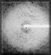
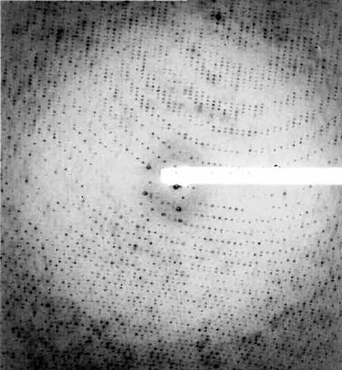
Diffraction pattern of a protein (Cytochorome C Oxidase) |
(1) Synchrotron radiation to study molecular structures
SR enables us to study an atomic level structure of a protein molecule. It is well known that proteins play very important roles in any life forms. Since the nature of proteins is originated fromtheir atomic structures, determination of atomic structure of proteins is one of the most important areas of study in biology.
The protein crystal scatters the SR according to its atomic structure resulting a very regular diffraction pattern (right). This pattern was obtained from a protein (Cytochrome C Oxidase) which plays an important role in the respiration mechanism.
The nutrition taken in by a living body is oxidized to generate energy. This protein works as a proton (Hydrogen ion) pump to create a gradient of proton concentrations across a cell membrane.
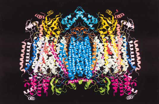
 Revealed fine structure of a Cytochrome C Oxidase from a cow’s heart (ribbon model representation) Revealed fine structure of a Cytochrome C Oxidase from a cow’s heart (ribbon model representation) |
Such a gradient is essential in the process of synthesizing adenosine 5’-triphosphate which is the most versatile forms of energy in cells. However, the mechanism of proton pumping is not fully understood at an atomic level.
A picture below shows a ribbon-model representation of a molecular structure of Cytochrome C Oxidase resulted from an analysis of almost five million diffraction spots in previous picture.
Such studies of proteins are widely appreciated owing to ontributions of synchrotron radiation and we have one of the best facilities for the structural study of macromolecules.
(2) Synchrotron radiation to study arrangement of electrons in crystals
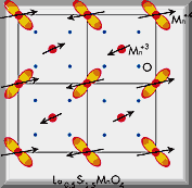
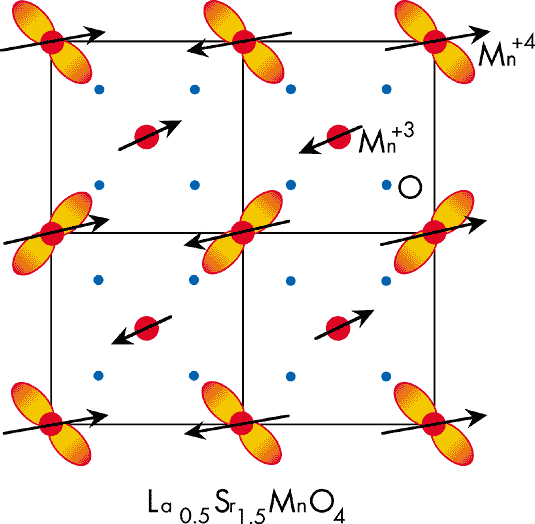
Charge and orbital ordering in a manganite crystal |
Some manganite crystals show drastic changes in electrical conductivity when exposed to a magnetic field. This henomenon draw much attention from scientists and engineers because it reflects correlation effects of electrons in solids and could lead to an invention of new magneticdevices.
Such effect is strongly related to a spatial ordering of charge, spin and orbit of electrons. Neutron diffraction technique enables to observe the spin ordering directly, but not the charge and orbital orderings.
However, a successful observation of the charge and orbital ordering of a manganite was made here for the first time using the resonant effect of the SR. The result of such studies also timulated theoretical discussions on resonant X-ray scattering.
(3) Synchrotron radiation to study soft biological tissues
X-ray images such as taken at hospitals show difference in absorbing power of different parts
of your body. Another way to see internal structure of a living body (without cutting open your
body) is making use of the difference in traveling speed of X-rays (phase contrast) through different parts of body. In light element materials, the phase contrast is thousands of times more sensitive than the contrast resulted from a difference in absorption so that we can see internal
structure much clearer.
A collaborative team of Hitachi Ltd., the PF and Tsukuba University developed a
computational tomographic imaging method based on the phase contrast of X-rays utilizing a
special type of X-ray interferometer. Figures below show a brain of a rat observed through two
techniques. Comparing the pictures one can see a clear superiority of the phase contrast technique
over the conventioal method.
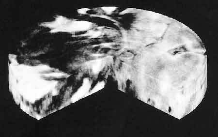
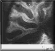
The superiority was also demonstrated when a liver of a rabbit was observed (bottom picture). A cancer in the liver was clearly visible with the phase contrast technique, but would be hard to spot when conventional method is employed. The research group is now trying to develop a new clinical imaging system using this method.
 |
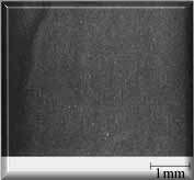 |
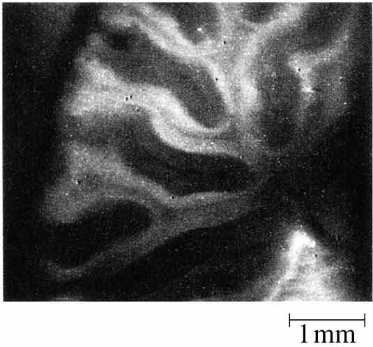
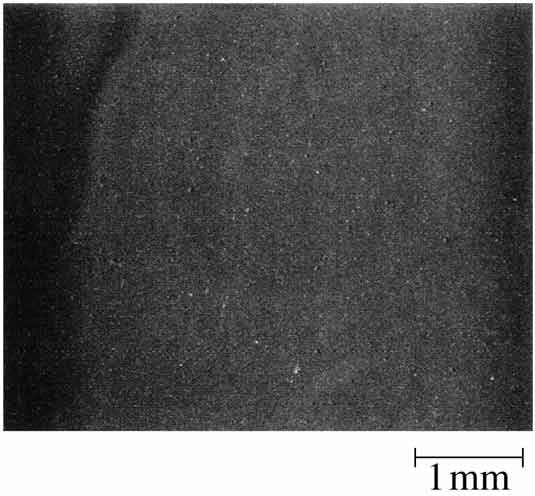
| Cross-sectional image of a rat’s brain |
| (a) Phase contrast method |
(b) Absorption contrast method |
 |
Phase contrast image of a rabbit’s liver.Dark area indicates liver cancer |
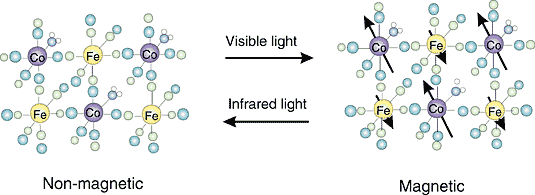
(4) Synchrotron radiation to study a molecule-based magnet
Magnetic materials are widely used for recording as in tape recorders where changing magnetic field controls the amount of magnetization on the tape. It is known that an electrical process induced by a light irradiation can also control magnetic properties of some materials (optical switching). A group of metal cyanide complexes has attracted a great interest because light irradiation converts their magnetic property quite easily: visible light converts the material to a ferromagnetic state while infrared light converts back to a non-magnetic state in a reversible way.
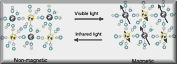 |
Optical switching of a olecule-based magnet Iron (Fe) and Cobalt (Co) atoms in Na0.4FeCo1.3(CN)6 molecules are magnetized by visible light and are demagnetized by infrared light. Arrows in the right figure show magnitudes of magnetization of Fe and Co atoms. |
However, the detailed mechanism has not been elucidated yet. X-ray absorption spectra of one of such materials was studied. In the spectra, there exists oscillatory structures called an X-ray Absorption Fine Structure (XAFS) which is a result of interference between emitted photo-electron wave and back-scattered photo-electron wave from neighboring atoms. This structure reflects chemical states of absorbing elements and distances to the neighboring atoms.
By analyzing XAFS spectra, the efficiency of light switching was quantitatively estimated.
Furthermore, detailed atomic structure change around metal atoms was quantitatively determined.
Such information is very helpful in the further development of new molecule-based magnetic
materials.
Radiation sources and beam handling techniques
Continuous efforts have been put in to improve this facility for all sorts of applications.Followings are some examples of improvements we have accomplished in recent years.
Radiation source
Continuous efforts have been made in developing and improving the SR source since its initialoperation in 1982. The recent, most notable accomplishment in this area was the emittanceupgrade of the 2.5 GeV ring in 1997-1998 after 4 years of preparation and construction of necessarycomponents of the ring. With the improvement, the electron beam size was made much smallerthan before (approx. 280 µm (horizontal) x 80 µm (vertical) ) and the brilliance of the ring wasincreased by a factor of approximately 5.
Insertion Devices
For even higher radiation intensity as well as to control polarization of the SR beam, severaltypes of Insertion Devices were developed and installed. They have many arrays of alternatingmagnetic field to force the electron beam to spatially oscillate. SR is emitted at every flip of themagnetic field when electron beams pass through. The SR overlaps with each other, generatingmuch more intense radiation. There are 6 insertion devices in the 2.5 GeV ring and 2 in the 6.5 GeVring. Two major technical contributions from the PF Insertion Device Group are : (1) InsertionDevices for generating circularly-polarized soft-X-rays or elliptically polarized X-rays by makingelectron beam travel along a spirally shaped trajectory and (2) in-vacuum undulater which isuseful for generating X-rays several orders of magnitude brighter than that from bending magnets.
SR beam handling technique
It is also important to efficiently transport synchrotron radiation to the sample position for experiments since we would otherwise waste the intense photon beam before it reaches the experimental station. Transporting the SR is done through a beamline that consists of many optical elements and beam transport pipes. Our PF Optics Group developed a transmission type diamond crystal X-ray phase plate that can control polarization state of X-rays very efficiently. Such phase plates are so efficient that they are now widely used at many synchrotron radiation facilities worldwide
Users of our Synchrotron Radiation Facility
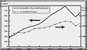 |
Activities per year
(dip in 1996~97 : reduced beam time due to a major upgrading work) |
700-800 experimental proposals are submitted to Program Advisory Committee and reviewed annually. Approximately 500 scientific papers are published and 30-40 doctoral theses are written every year from experiments carried out at our facility.
We are open to outside institutions to construct their own beamlines at their costs. Organizations which have built and are operating such beamlines are the Australian Nuclear Science and Technology Organization (see next article in this issue) , 4 industrial companies (NTT, NEC, Hitachi and Fujitsu), Institute of Solid State Physics of the University of Tokyo, Spectrochemistry Laboratory of the University of Tokyo and Tsukuba Advanced Research Alliance of Tsukuba University. We are one of the most active centers for SR research and any users from any countries are most welcome. Professors of the Photon Factory have joint appointments with the Graduate University of Advanced Studies so that a number of graduate students and post-doctoral fellows are studying and working on synchrotron-radiation-related science. Any students who have interests and enthusiasm are most welcome.