|
|
|
Early Detection of Breast Cancer
A novel refraction-based X-ray imaging for medical diagnosis was developed. Ordinary absorption-based X-ray imaging, MRI (magnetic resonance imaging), and sonography can detect a cancer greater than 5mm. Refraction-based X-ray imaging, utilizing a synchrotron radiation could become a powerful tool for early cancer diagnosis. As the synchrotron radiation has the penetration power like a laser light, one can construct an X-ray optics called X-ray dark-field imaging. We use a crystal monochro-collimator to make incident X-rays going straight forward and analyzer crystal angular filter to filtrate refracted X-ray component. The long history of siliconbased electronics industry made near defectless silicon crystals available to us. A part of a body is placed in between them in a future. A silicon angular filter with a certain thickness may have zero transmission for the straight component of light that does not have received refraction through the specimen while refraction component can pass through this angular filter.
We can survey 90mm x 90mm field in one shot using 35 keV X-rays. An excised human femoral head shows an articular cartilage that has not been visualized by any other technique. This research work was awarded a prize by the Society of Applied Physics of Japan When this technique was applied to a breast cancer tissue, 2-dimensional distribution of breast cancer nests was revealed with extremely high contrast and high spatial resolution of 30 microns.
(picture below) We achieved spatial resolution of approx. ~10microns so that each cancer cell could be seen. This will lead to clinical diagnosis and pathological diagnosis. Thanks to the higher penetration power of the 35 keV X-ray, patients will go through less painful preparation process.
In a future we would like to achieve the image reconstruction for not only breast but also other organs such as lung.(Figure on page 10)

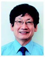
The author of this article, Dr. Masami Ando is a Professor of KEK as well as of Graduate University of Advanced Studies. He developed medical imaging techniques for clinical diagnosis using synchrotron radiation. His hobby is on butterfly and railway.
|
|
