|
|
|
Protein structure analysis using synchrotron X-ray beam lines
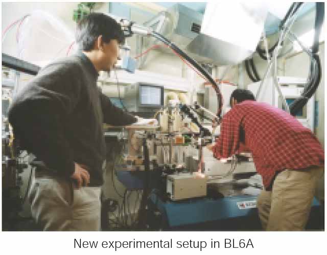
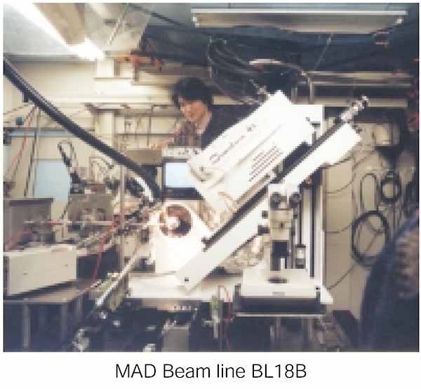
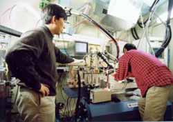
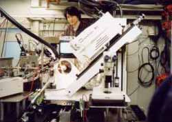
Three dimensional arrangements of proteins and large protein complexes can be studied in atomic detail by synchrotron X-ray crystallographic techniques. In the era of third-generation synchrotron facilities specializing in insertion device beam lines, the second generation facilities such as the PF face severe competitions. The protein crystallography beam lines in operation at the PF, BL6A (below left), BL6B, and BL18B (below right), remain very productive; 40 to 70 original structural papers are published each year. In fact, 80 to 90% of protein crystallography experiments can be performed on beam lines at any well set-up and maintained second generation synchrotron facilities. It is thus critical for the PF to keep developing new technologies for protein crystallography experiments and implement them in the facility. We are vigorously working on this by (1) constructing new insertion-device beam lines on the 6.5 GeV Advanced Ring (AR) and the 2.5 GeV PF ring, and (2) developing key technologies for high-throughput protein crystallography experi-ments. They include a unified database for entire experiments (developed by Y. Gaponov), automatic capturing of protein crystals and cryogenic freezing, and a next generation X-ray area detector. For instance, the new experimental table and the singlebounce monochromator allows for anomalous experiments on BL6A (page 5 bottom left). The energy resolution of the monochromator is not ideal for multiple anomalous dispersion (MAD, a technique to determine protein structures rapidly and accurately using anomalous scattering effects of heavy atoms embedded in protein crystals) experiments, but the energy tuneability has improved the success rate of structure determination on the beam line. Indeed, we have already solved a structure of a domain of a protein involved in the vesicle transport using the MAD technique on BL6A (page 5, bottom right).
 |
|
|
New experimental setup in BL6A |
MAD Beam line BL 18B
|

|
The first structure determined of a transport protein from a Se-MAD experiment using the new set-up of BL6A (T. Nogi et al., submitted to Nature Structural Biology).
|
Furthermore, a new section with an experimental floor, 1279 m2, is soon to be completed along the northwest quadrant of the 6.5 GeV AR where we will construct a new MAD beam line. In collaboration with Profs. H. Kawata, M. Nomura, and Assoc. Prof. S. Yamamoto of the PF, the Structural Biology group has designed the beam line optics, hutches and experimental facilities as well as the adjacent preparation laboratory. The beam line will be constructed on one of the straight sections, NW12. A 40- mm period undulator will produce X-ray photons ranging from 7 to 16.8 KeV. The flux at the sample position is expected to be 2×10¹² photons/sec through an aperture of 200 µm by 200 µm, which will surpass the intensity of bending magnet beam lines of the third generation synchrotron sources. As a next phase, we have a plan to build new insertion device MAD beam lines on the PF-Ring and are making vigorous efforts for realization of the new beam lines along with the reorganization of the PF-Ring to create more straight sections.
|
|