Photoelectron diffraction measurements of gaseous molecules aligned in one direction: towards ultrafast molecular imaging
PressreleaseOctober 16, 2015
High Energy Accelerator Research Organization (KEK)
RIKEN
Japan Synchrotron Radiation Research Institute (JASRI)
Researchers from High Energy Accelerator Research Organization (KEK), the University of Tokyo, Ritsumeikan University, Chiba University, Kyoto University, Japan Atomic Energy Agency, RIKEN, and Japan Synchrotron Radiation Research Institute (JASRI) have successfully observed the X-ray photoelectron diffraction pattern of gaseous iodine molecules (I2) aligned in one direction by using the X-ray free electron laser (XFEL) at the SACLA facility in Harima, Japan.
The results were published in the September 15 issue of the online scientific journal Scientific Reports.
Photochemical reactions, which cause structural changes in molecules under light irradiation, have been applied to optical switches and optical memory devices. However, the mechanism has not been fully clarified because the reactions occur on ultrafast time scales of femtoseconds to picoseconds. Thus, the research group are developing a molecular imaging method (molecular movie), based on photoelectron diffraction (Fig. 1). The method will be applied to the study of ultrafast photochemical reaction dynamics and methods of reaction control. The creation of a molecular movie that visualizes the ultimate reaction in both time and space is currently a topic receiving close review and has stimulated the interest of molecular scientists worldwide.
 Fig. 1 Principle of photoelectron diffraction
(Left) Photoelectron wave emitted from the 2p inner orbital of the left iodine atom (I)
(Center) Scattered wave produced when the photoelectron is scattered by the right iodine atom (I)
(Right) Interference pattern produced by superposition of the two waves
The atomic distance is determined from the interference pattern.
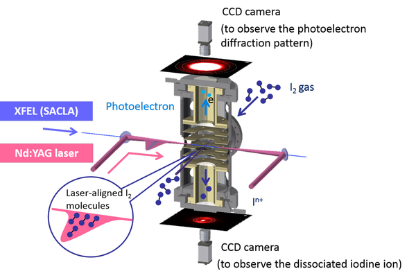
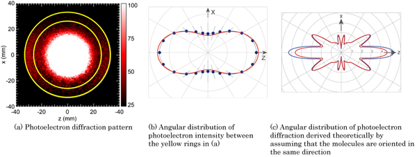
Because gaseous molecules are oriented randomly and their photoelectron diffraction patterns are averaged, the photoelectron diffraction pattern of a particular molecule is not obtainable in conventional experiments. Therefore, in this experiment, gaseous iodine molecules (I2) are irradiated with YAG laser pulses so as to align them by an electric field of the laser (Fig. 2). Thus, the photoelectron diffraction pattern of the I2, Fig. 3 a, is successfully obtained by irradiating it with XFEL pulses, which are synchronized in time and space with the YAG laser pulses.
After emitting a photoelectron, the I2 dissociates into iodine ions (In+), which are emitted in the direction of molecular orientation. The degree of alignment of the I2 is determined by the measurements of the iodine ions.
The experimental results of the photoelectron diffraction pattern are consistent with the theoretical calculations (Fig. 3b, red line) performed by considering the degree of alignment.
On the other hand, the theoretical calculation that assumes the molecules are completely aligned with each other predicts the appearance of an interference pattern in the photoelectron diffraction pattern (Fig. 3c). However, in the experiments, an interference pattern was not observed owing to the limitations in molecular alignment. A "molecule movie" that visualizes changes of the molecular structure during an ultrafast photochemical molecular reaction will be able to be realized by observing interference patterns through a better alignment of the molecules.
 Fig. 2 Basic concept of the photoelectron diffractmeter
In the pulsed molecular beam, iodine molecules are aligned in the direction of the electric field of the Nd:YAG laser pulse. When photoelectrons are emitted from the iodine molecules after exposure to the XFEL pulse synchronized with the Nd:YAG laser pulse, the molecular bond breaks and the iodine atom decomposes into iodine ions. The speed distribution of the photoelectrons is observed through the upper CCD, while the speed distribution of the dissociated ions is observed through the lower CCD.
 Fig. 3 Photoelectron diffraction pattern and its angular distribution
(a) Two-dimensional diffraction pattern of photoelectrons. The 2p photoelectrons of an iodine atom (I) are observed in the area between the yellow concentric circles. The figure shows that the high-intensity 2p photoelectrons are emitted in the z-axis direction (in this case, the horizontal direction), parallel to both the electric field vector of the XFEL beam and the molecular axis of the iodine molecule.
(b) Angular distribution of 2p photoelectrons in the xz-plane in polar coordinates. The intensity of photoelectrons is proportional to their distance from the center. The black dots represent the experimental results and the red curve shows the calculation results when changes in the molecule's orientation are considered.
(c)Theoretically calculated angular distribution of 2p photoelectrons in the xz-plane in polar coordinates. In this calculation, the interference between the photoelectron wave and scattered wave is prominent because all molecules are oriented in the z-axis direction. The red curve shows the calculation results with one-time scattering, while the blue curve represents the calculation results with infinite-time scattering.
|
|
Media Contact
[ for research ]
Akira Yagishita
Senior Fellow, Institute of Materials Structure Science, High Energy Accelerator Research Organization (KEK)
E-mail: akira.yagishita@kek.jp
[ for public relations ]
Saeko Okada
Public Relations Office, High Energy Accelerator Research Organization (KEK)
TEL: +81-29-879-6047
FAX: +81-29-879-6049
E-mail: press@kek.jp
Related Link
Institute of Materials Structure Science, KEK
Photon Factory
詳しくは下記のページをご覧ください
http://