A research group composed of Dr. Naoki Sunaguchi (Gunma University), Prof. Tetsuya Yuasa (Yamagata University), M.D. Rajiv Gupta (Massachusetts General Hospital), Shin-ichi Hirano (Mercian Cleantec Corporation, MiZ Company Limited), and Prof. Masami Ando (Tokyo University of Science and Emeritus Professor at KEK) developed a new algorithm to improve the quality of an X-ray phase-contrast image.
X-ray phase-contrast imaging can provide far higher contrast in soft tissue compared to classical absorption-based imaging. Many groups have been developing a variety of imaging methods for potential clinical use. All these imaging methods suffer from a common problem: severe imaging artefacts arise when x-ray phase alternation exceeds the dynamic range of the imaging system, typically in the vicinity of bones and dense calcifications. These artefacts are similar to the metal and beam-hardening artefacts seen in traditional attenuation-based X-ray computed tomography (CT) even though they tend to be more severe and have a different physical basis. A particularly worrisome part of this type of artifact is the fact that it spreads broadly across a wide area on CT image even when the dense tissue responsible for it localized.
In order to suppress this unwanted artefact, the research team developed and tested a novel artefact suppression algorithm for x-ray phase-contrast computed tomography. As a first step, their algorithm identifies the regions in the image that exceed the dynamic range of the phase image based on the reconstructed absorption image. Identified region is mapped on the projection dataset and is treated as missing information during reconstruction. They use an iterative reconstruction algorithm that goes back and forth between the projection data set and phase image while imposing a priori information on both.
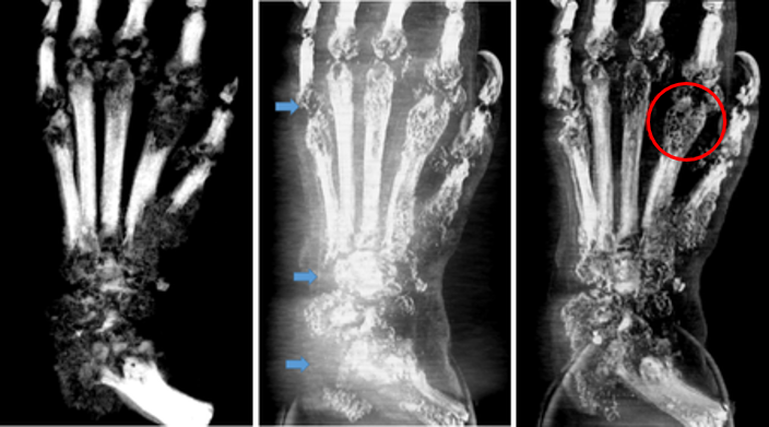
The proposed algorithm is quite effective. When tested in three different specimens --- iliac and coronary artery specimens containing atherosclerotic calcification, and a rat model of human rheumatoid arthritis ---the algorithm resulted in considerable reduction in dense structure artifacts. Figure below shows 3D Maximum Intensity Projection images of a rat foot modeling rheumatoid arthritis. As can be seen in the regions marked by arrows, there is near complete signal drop out due to artefacts that are dramatically improved by the proposed algorithm.
A large body of current research focuses on metal artifact reduction in conventional absorption-based CT for radiation therapy and quantitative diagnosis. The proposed algorithm will have direct application for this task. It will also be beneficial in future when phase-contrast imaging is clinically deployed.
This research was published in PLOS ONE on August 21 14:00 (the Eastern Standard Time of U.S.). Part of the experiment was performed under the S2 type proposal (project-type study) approved by the Photon Factory Program Advisory Committee (2008S2-002).
Title: "In vitro validation of an artefact suppression algorithm in x-ray phase-contrast computed tomography" [ DOI:10.1371/journal.pone.0135654 ]
Authors: Naoki Sunaguchi, Tetsuya Yuasa, Shin-ichi Hirano, Rajiv Gupta, Masami Ando